
The Role of Digital Scanning in Modern Orthodontics
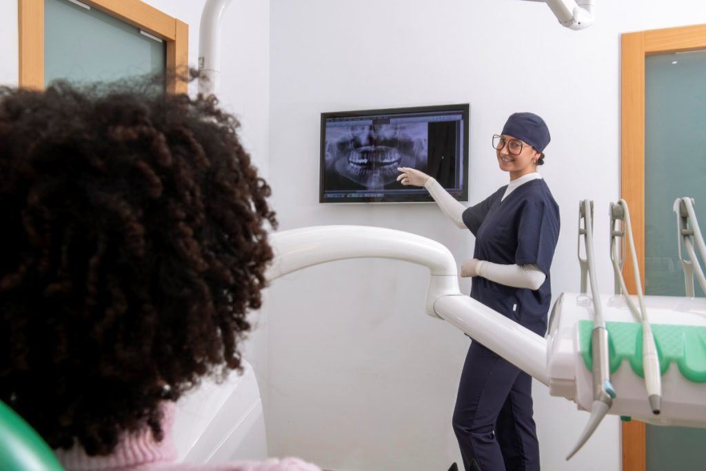
Anyone who had braces in the 1990s or 2000s remembers the impressions. The metal trays. The thick, cold alginate paste. The fifteen seconds of breathing carefully through your nose while it set, hoping you wouldn’t gag. For decades, this was simply how orthodontic treatment started, and patients accepted the discomfort because there was no alternative.
That entire process is now obsolete. With the introduction of technology in modern orthodontics, a digital intraoral scan takes a few minutes, produces no discomfort whatsoever, and generates data that’s substantially more accurate than the impression material it replaced. The change has been one of the most genuinely transformative shifts in orthodontic practice this century, and it affects everything from diagnosis to treatment planning to the speed and predictability of results.
What Actually Happens During a Digital Scan?
The clinician passes a small, wand-shaped scanner around the surfaces of your teeth, gums, and bite. The device captures thousands of images per second and stitches them together into a precise three-dimensional model of your mouth. The whole process typically takes between three and five minutes, and most patients are surprised by how unintrusive it feels.
The scanner uses structured light, optical sensors, or a combination of technologies depending on the manufacturer. iTero and 3Shape Trios are the two systems most commonly used in UK orthodontic practice, both producing exceptionally detailed digital impressions that surpass what physical alginate could ever capture.
There’s no waiting for material to set, no need to retake the scan if a section comes out poorly (the clinician simply rescans the area in question), and no shipping a physical model to a lab. The data exists immediately and can be sent to wherever it needs to go in seconds.
Why Accuracy Matters More Than You’d Think
Traditional impressions captured a reasonable approximation of tooth and jaw geometry, but they had real limitations. Material distortion during setting, dimensional changes during shipping, and translation errors when stone models were poured from the impressions all introduced small inaccuracies. Most of the time, those inaccuracies didn’t matter clinically. Sometimes they did.
Digital scans eliminate this entire chain of potential error. The data is captured at submicron accuracy and remains perfectly stable, with no physical degradation between capture and use. For complex orthodontic cases, particularly those involving precise tooth movements or surgical planning, this accuracy translates directly into better outcomes.
A few millimetres of error in an impression doesn’t sound like much. In a treatment plan that involves moving teeth into specific final positions, those millimetres compound into visible differences in the finished smile.
How Digital Scans Transform Treatment Planning
Once your mouth exists as digital data, the planning possibilities expand considerably. Treatment software can simulate tooth movements before any physical brace or aligner is fitted, predicting how teeth will respond to specific forces over specific timelines. You can see your projected outcome at the consultation stage, before committing to anything.
Invisalign’s ClinCheck system is the most familiar example. Your scan feeds directly into software that maps the entire planned course of treatment, aligner by aligner, showing you exactly how your teeth will move from their current position to the final result. You see your future smile as a 3D model before you’ve decided whether to proceed.
For fixed braces, digital data enables custom bracket placement using indirect bonding techniques. Brackets can be positioned with sub-millimetre precision in the digital plan, then transferred to your teeth using a custom tray that places each one exactly where the planning software determined it should sit. The clinical accuracy is significantly higher than freehand placement.
The Speed Implications
Beyond the patient comfort element, digital workflows are simply faster than analogue ones at every stage. A consultation that previously required scheduling a separate impression appointment can now happen in a single visit. Treatment planning that took weeks of back-and-forth between clinic and lab now happens in days. Aligner manufacturing that once required physical models being shipped internationally now happens directly from the digital file.
For the patient, this often means treatment starts sooner. A scan taken on Monday could result in aligners arriving within two to three weeks, rather than the four to six weeks that physical impressions previously required.
Monitoring Progress with Digital Tools

Digital scanning doesn’t stop being useful once treatment begins. Periodic rescans throughout the course of treatment let the clinician compare your current position to the planned trajectory and identify any deviations early. Some practices use overlay software that shows your actual progress against the predicted progress, with deviations colour-coded for immediate attention.
This kind of monitoring used to be impossible. You couldn’t easily compare a current physical impression to a previous one in any meaningful way. Now, comparison is automatic and precise, which means course corrections happen sooner and treatment stays on track more reliably.
What This Means for Patients
The clinical benefits aside, digital scanning has changed the experience of orthodontic treatment in ways that matter for patients who’d otherwise hesitate.
Patients with strong gag reflexes who previously couldn’t tolerate impressions can now have orthodontic treatment without that obstacle. Children who’d find traditional impressions distressing find scans much easier to tolerate. Anyone with a busy schedule benefits from the time saved at every stage.
Beyond comfort, the visualisation tools create a different kind of conversation between clinician and patient. Seeing your planned outcome in three dimensions before treatment begins makes the decision-making process more concrete, and engagement with treatment plans tends to be higher when patients can see exactly where they’re heading.
The Limits of the Technology
Worth being honest about what digital scanning doesn’t do. It doesn’t replace clinical expertise; the scanner produces data, but interpreting that data and designing the right treatment plan still requires an experienced orthodontist. The technology is a tool, not a substitute for skill.
It also doesn’t perform miracles on every case. Complex orthodontic problems still require careful planning, time, and patient compliance regardless of how accurate the initial scan was. Digital workflows make good orthodontists faster and more precise; they don’t make all orthodontists equally good.
Modern Practice Today
Boston Orthodontics has been using digital scanning workflows since they became clinically reliable, and our experience has been that the technology genuinely changes outcomes for the better. Faster diagnosis, more accurate planning, more predictable treatment, more comfortable patient experience. There aren’t many shifts in clinical practice that improve every dimension at once, and digital scanning has been one of them.
If you’re considering treatment and want to see what your projected outcome looks like before deciding, an initial consultation with an orthodontist in London for teeth straightening treatments at our Belgravia clinic includes a digital scan and a clear visualisation of what your treatment would look like.
Book Your Dental Checkup Today
Ready to take control of your oral health? Whether it’s your first visit or a routine cleaning, regular dental checkups make all the difference. Our expert team is here to help you maintain a strong, confident smile.
MOST POPULAR TREATMENTS
Our Services

Our Doctor
Meet Doctors

Dr. Elif Keser
Dentist

Dr. Tania Rafael Garcia
Dental Surgeon

Dr. Gustavo Silva
Dental Surgeon
